IBA Dosimetry
PhantomX Head/Neck Phantoms
Art. N°
Hover to zoom · click to enlarge
Medical Imaging - QA for AI
CTA
Advanced head and neck phantoms designed to simulate true anatomical detail and vascular structures, elevating your imaging performance and AI-driven diagnostics.
- Brochure
- /fileadmin/user_upload/products/09_phantomx/PhantomX_-_Head_Phantom/IBA-PhantomX-HeadNeckPhantom-Brochure-Working.pdf
- Specifications
- Specifications Size: Approx. 185 x 215 x 322 mm Weight: Approx. 5630 g Base material: Cellulose-polymer composite Optimal tube voltage: 120 kVp – adaptable upon request –
- Used for Workflow
- Head, Neck
- Diagnostic features
- Diagnostic features Realistic simulation of head and neck vessels up to the aortic arch, bone and soft tissues. Calcifications of the internal carotid artery at the carotid bifurcation on both sides with mild stenosis on the right side.
- Packaging & Dimensions
- Packaging & Dimensions The product ships in a protective hard case featuring interior foam padding. - Dimensions (case): 520 x 350 x 220 mm - Dimensions (packaged)*: 572 x 425 x 239 mm - Total weight (ready to ship): 12.0 kg *Outer packaging (e.g., cardboard box or protective wrap) may vary slightly, affecting total dimensions.
- Head / Neck Phantom CTA
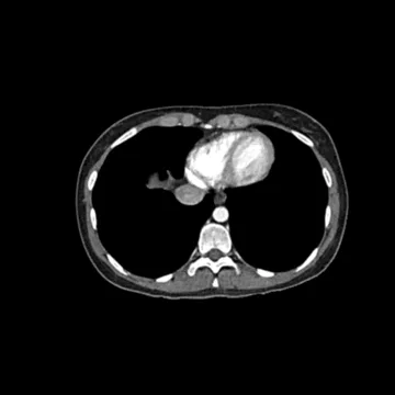
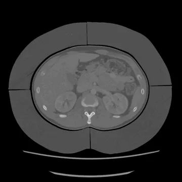
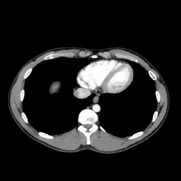
- This phantom simulates a contrast-enhanced head in the arterial phase (CT angiography), including the neck and upper thorax up to the aortic arch and the fifth thoracic vertebra. The vertex is included to approximately 0.5 cm below the skin level. The thoracic section excludes shoulder and back parts that are typically not part of CTA examinations. The internal carotid arteries feature bilateral calcifications, with moderate stenosis on the right side, while no other significant vascular pathologies are present. The phantom provides a detailed and realistic representation of vascular structures, soft tissue, and bone, including fine anatomical details such as lymph nodes. Air voids are filled with a cellulose-polymer composite with an attenuation of approximately −160 HU. It can be used in CT (including CBCT) to evaluate and optimize CTA imaging performance and post-processing applications such as vessel segmentation, including AI-enabled solutions, and is also well suited for training purposes.
- Compatible with Machine/Modality/Application
- Head, Neck, CT Simulation, CBCT